Smartalpha

logo-smartalpha

Anesthesia &

Pain Medicine

Owning this surprising bit of technology at your clinic will make sure that you have the most unique regional anesthesiology experience to date.

Owning this surprising bit of technology at your clinic will make sure that you have the most unique regional anesthesiology experience to date.

Nerveblox is a CE-marked, AI-enabled ultrasound guidance software to assist anesthesiologists with sonoanatomy.

nerveblox-ai

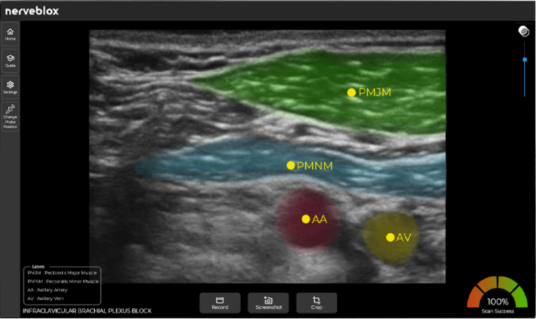
Nerveblox makes ultrasound images colorful and easily interpretable. The AI at the heart of it gives anesthesiologists extra confidence while performing ultrasound-guided peripheral nerve blocks. It assists physicians in quickly interpreting ultrasound images by auto-labeling decisive anatomical landmarks at each block region.

Regional anesthesia is transforming in time thanks to advancements in technology.

PNB with theNerve Stimulator US-guided PNB ParesthesiaTechnique Artificial Intelligenceguided PNB 1950s 1980s TODAY 1990s

In the beginning, physicians were performing basic anatomical techniques. The variety of block types was limited, and the associated complication rates were arguable.

Then, an electric stimulator was introduced to localize the nerve more accurately which definitely increased block success and the number of accepted block types.

Within the last decade and with the increasing accessibility of ultrasound systems, ultrasound-guided peripheral nerve blocking emerged and resulted in fewer complications and broader use-cases than its predecessors.

Finally, the age of artificial intelligence-guided nerve blocks has arrived.

What is Nerveblox?

Nerveblox is an AI-driven decision support solution to help anesthesiologists practice peripheral nerve block (PNB) faster.

Nerveblox is able to capture real-time images from any ultrasonography (US) device. The captured images are then processed by state-of-the-art artificial intelligence algorithms, and the key anatomical landmarks are automatically labelled for the user. All you have to do is plug Nerveblox into any compatible ultrasound device.

Today, Nerveblox supports the following peripheral nerve block (PNB) types:

The interscalene approach to brachial plexus block results in reliable anesthesia of the shoulder and upper arm. Interscalene block anatomy landmarks are as follows:

• Brachial Plexus
• Sternocleidomastoid Muscle
• Anterior Scalene Muscle
• Middle Scalene Muscle
The supraclavicular approach to the brachial plexus block results in anesthesia of the upper limb including often the shoulder because all trunks and divisions can be anesthetized from this location. Supraclavicular block anatomy landmarks are as follows:

• First Rib
• Parietal Pleura
• Subclavian Artery
• Brachial Plexus
The infraclavicular block provides a block of the arm below the shoulder. Infraclavicular block anatomy landmarks are as follows:

• Pectoralis Major Muscle
• Pectoralis Minor Muscle
• Axillary Artery
• Axillary Vein
The superficial cervical plexus nerve block results in anesthesia of the skin of the anterolateral neck and the ante-auricular and retro-auricular areas, as well as the skin, overlying and immediately inferior to the clavicle on the chest wall. Superficial block anatomy landmarks are as follows:

• Carotid Artery
• Sternocleidomastoid Muscle
• Anterior Scalene Muscle
• Cervical Plexus
• Jugular Vein
The axillary brachial plexus block (including the musculocutaneous nerve) results in anesthesia of the upper limb from the mid-arm down to and including the hand. Axillary block anatomy landmarks are as follows:

• Axillary Artery
• Musculocutaneous Nerve
• Biceps Brachii Muscle
• Conjoint Tendon
• Triceps Brachii Muscle
• Coracobrachialis Muscle
The pectoral nerve (Pecs) block I and II are a novel technique to block the pectoral nerves. These blocks can be used to provide analgesia for a variety of anterior thoracic wall surgeries, most commonly breast surgery. Pecs I & II block anatomy landmarks are as follows:

• Pectoralis Major Muscle
• Pectoralis Minor Muscle
• Serratus Anterior Muscles
• Parietal Pleura
• First Rib
nerveblox-user interface
Transversus abdominis plane (TAP) block is a regional technique for analgesia of the anterolateral abdominal wall. TAP block anatomy landmarks are as follows:

• Peritoneal Cavity
• Transverse Abdominis Muscle
• Internal Oblique Muscle
• External Oblique Muscle
The rectus sheath nerve block is a useful technique for umbilical surgery, particularly in pediatric patients. Rectus block anatomy landmarks are as follows:

• Rectus Sheath (Posterior Aspect)
• Rectus Abdominis Muscle
• Rectus Sheath (Anterior Aspect)
• Peritoneal Cavity
The ultrasound (US)-the guided technique femoral nerve block is used for the femur, patella, quadriceps tendon, and knee surgery, and analgesia for hip fracture. Femoral block anatomy landmarks are as follows:

• Femoral Artery
• Femoral Vein
• Femoral Nerve
• Iliopsoas Muscle
An Adductor Canal nerve block is useful as a sciatic nerve block for foot and ankle procedures. Adductor block anatomy landmarks are as follows:

• Saphenous Nerve
• Femoral Artery
• Sartorius Muscle
• Vastus Medialis Muscle
The popliteal fossa nerve block is a very clinically valuable technique that results in anesthesia of the calf, tibia, fibula, ankle, and foot. Popliteal block anatomy landmarks are as follows:

• Common Peroneal Nerve
• Tibial Nerve
• Popliteal Artery
• Popliteal Vein
Erector spinae plane (ESP) block is an interfascial plane block where a local anaesthetic is injected in a plane preferably below the erector spinae muscle. ESP block anatomy landmarks are as follows:

• Trapezius Muscle
• Rhomboid Muscle
• Erector Spinae Muscle
• Transverse Process
• Parietal Pleura