SmartAlpha’s Nerveblox™ receives FDA 510(k) clearance. Learn more
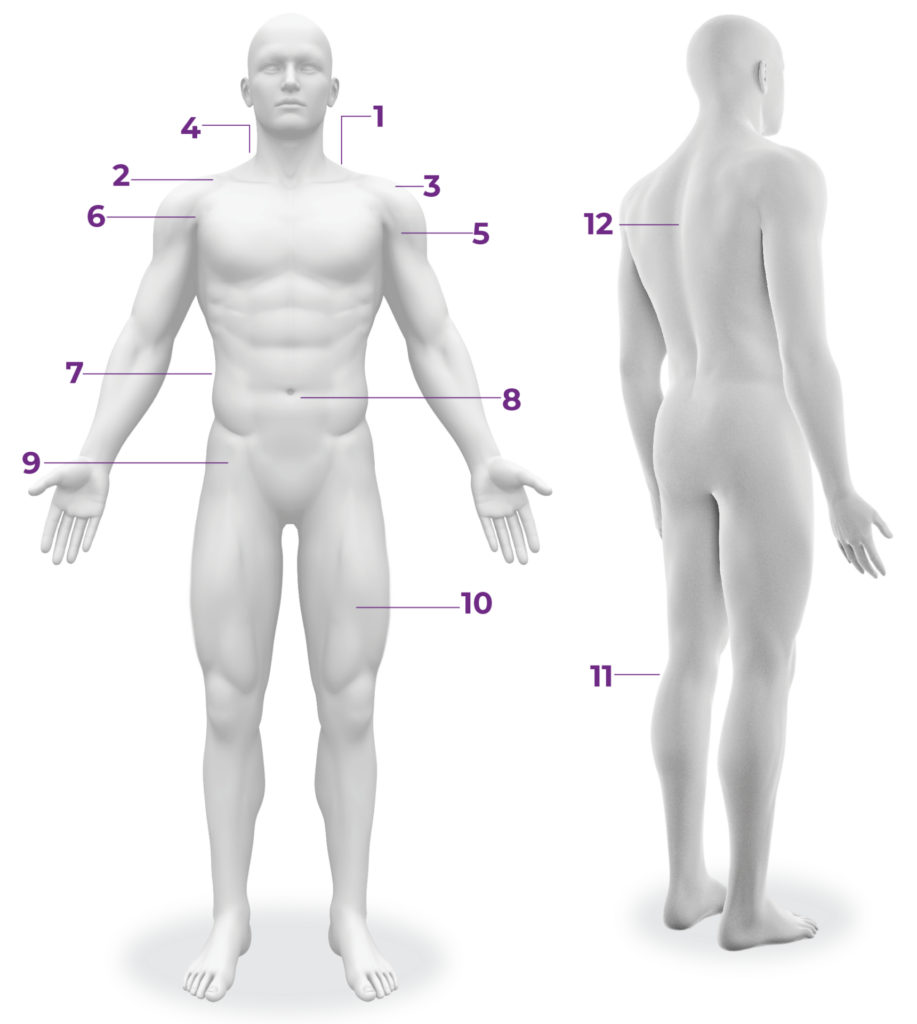
Nerveblox™ assists in the visualization of anatomical structures across 12 regional anesthesia procedures, with its real-time AI detecting 50+ anatomical structures.
Nerveblox™ assists in the visualization of anatomical structures across 12 regional anesthesia procedures, with its real-time AI detecting 50+ anatomical structures.
Nerveblox™ is an
FDA-cleared, CE-marked,
AI software solution that assists users with interpreting sonoanatomy in real-time.
BP:Brachial Plexus SA:Subclavian Artery
PL:Pleura FR:First Rib
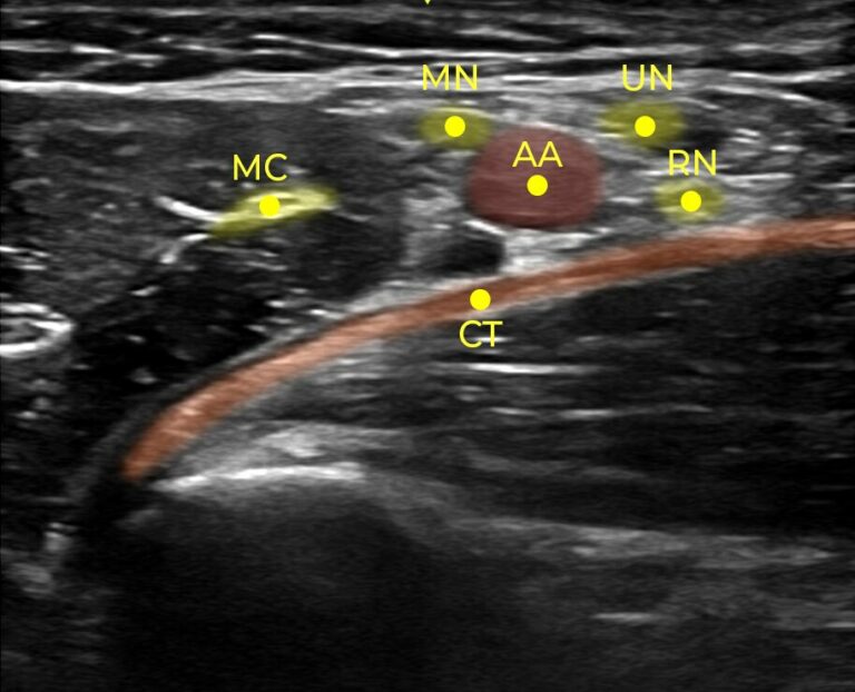
Nerveblox™ makes ultrasound images colorful and easily interpretable. The AI at the heart of it provides real-time guidance while performing ultrasound-guided peripheral nerve blocks. It assists physicians in interpreting ultrasound images by auto-labeling decisive anatomical landmarks for each block region.
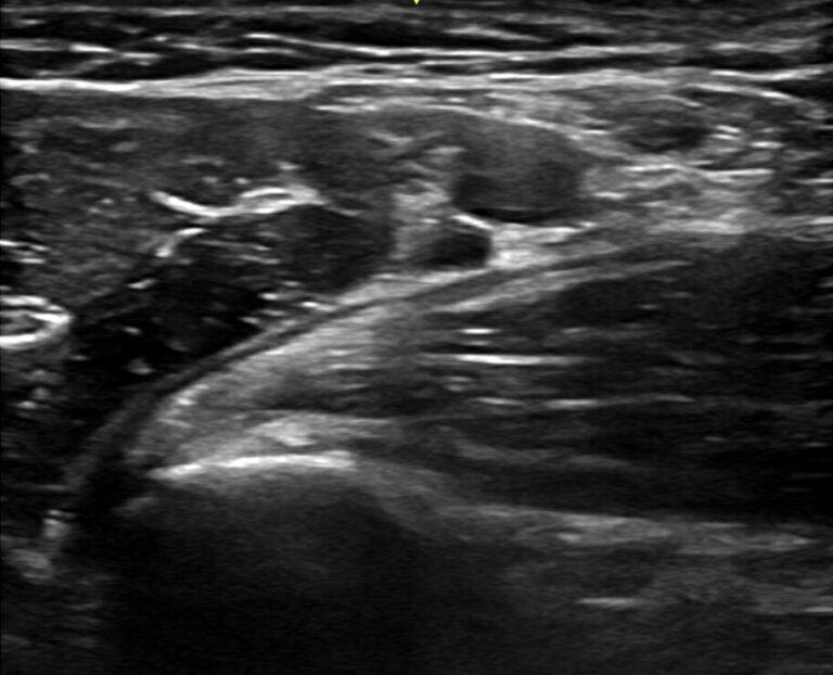
Traditional Ultrasound Image
Ultrasound image with NerveBlox™
Nerveblox™ supports 12 regional anesthesia procedures