SmartAlpha

SmartAlpha’s Nerveblox™ receives FDA 510(k) clearance. Learn more

logo-smartalpha

The Intelligence Layer for Ultrasound — for Human and Autonomous Healthcare Workers

Robotic Healthcare worker performing ultrasound scan using SmartAlpha AI

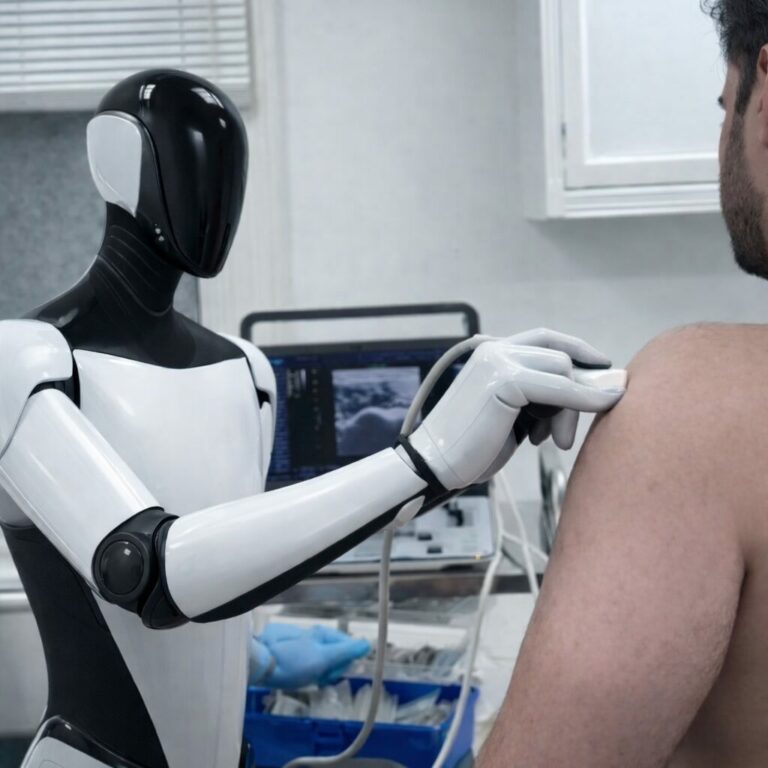
SmartAlpha solutions are built to power the next generation of AI-assisted healthcare workers, removing the skill barriers historically required for expert-level ultrasound scanning, whether performed by human clinicians or autonomous ultrasound users.

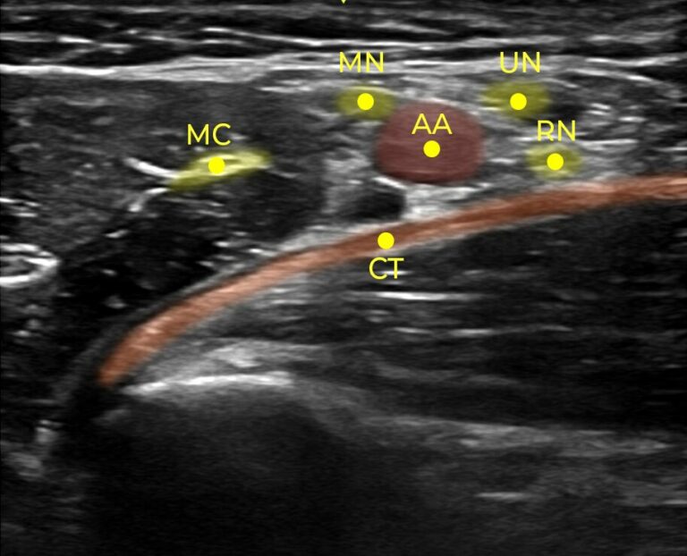
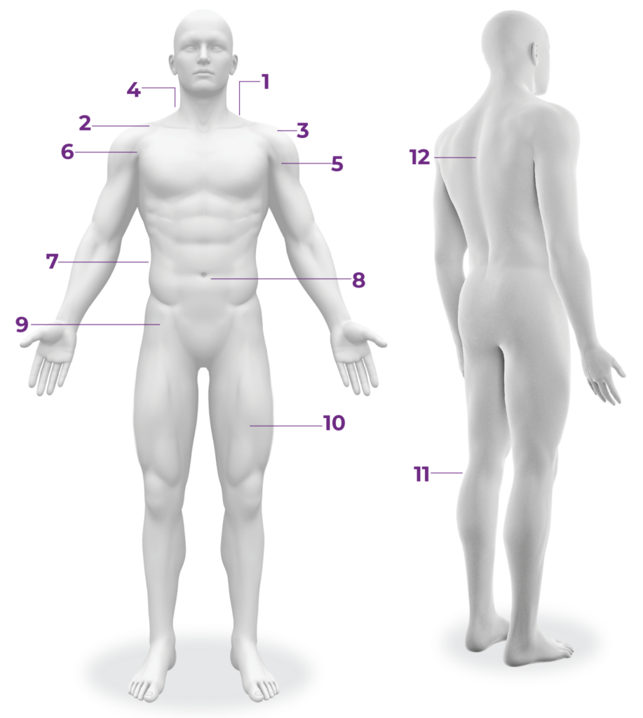
Today, the SmartAlpha solutions support 50+ scan procedures, 200+ anatomical structures, and performs real-time image optimality assessment required to guide hundreds of transducer manipulation strategies.

Solid vision… Moving at the speed of AI.

Available Solutions

Nerveblox™ assists in the visualization of anatomical structures across 12 ultrasound use cases exclusive to regional anesthesia, with its real-time AI detecting 50+ anatomical structures.

Nerveblox™ assists in the visualization of anatomical structures across 12 regional anesthesia procedures, with its real-time AI detecting 50+ anatomical structures.

Nerveblox™ is an FDA-cleared, CE-marked, AI software solution that assists users with ultrasound scanning for regional anesthesia.

In the above scan, SmartAlpha AI identifies and tracks several structures including the Pleura (PL) and the 1st Rib (FR) which are the most critical landmarks that should be visualized during scanning.

Enhances ultrasound image interpretation during live scanning.

Key anatomical structures are automatically identified in real time, providing immediate anatomical awareness to the user.

An integrated image quality indicator continuously evaluates anatomical visibility and completeness, providing feedback to the user.

Traditional Ultrasound Image

Ultrasound image with SmartAlpha AI

Today, Nerveblox™ supports 12 ultrasound procedures

nerveblox-user interface
Transversus abdominis plane (TAP) block is a regional technique for analgesia of the anterolateral abdominal wall. TAP block anatomy landmarks are as follows:

• Transverse Abdominis Muscle
• Internal Oblique Muscle
• External Oblique Muscle
• Peritoneal Cavity
The rectus sheath nerve block is a useful technique for umbilical surgery, particularly in pediatric patients. Rectus block anatomy landmarks are as follows:

• Rectus Abdominis Muscle
• Peritoneal Cavity
• Rectus Sheath (Anterior Aspect)
• Rectus Sheath (Posterior Aspect)
The ultrasound (US)-the guided technique femoral nerve block is used for the femur, patella, quadriceps tendon, and knee surgery, and analgesia for hip fracture. Femoral block anatomy landmarks are as follows:

• Femoral Vein
• Femoral Nerve
• Femoral Artery
• Iliopsoas Muscle
An Adductor Canal nerve block is useful as a sciatic nerve block for foot and ankle procedures. Adductor block anatomy landmarks are as follows:

• Femoral Artery
• Sartorius Muscle
• Vastus Medialis Muscle
• Saphenous Nerve
The popliteal fossa nerve block is a very clinically valuable technique that results in anesthesia of the calf, tibia, fibula, ankle, and foot. Popliteal block anatomy landmarks are as follows:

• Common Peroneal Nerve
• Tibial Nerve
• Popliteal Artery
• Popliteal Vein
Erector spinae plane (ESP) block is an interfascial plane block where a local anaesthetic is injected in a plane preferably below the erector spinae muscle. ESP block anatomy landmarks are as follows:

• Trapezius Muscle
• Erector Spinae Muscle
• Transverse Process
• Pleura
The interscalene approach to brachial plexus block results in reliable anesthesia of the shoulder and upper arm. Interscalene block anatomy landmarks are as follows:

• Brachial Plexus
• Anterior Scalene Muscle
• Middle Scalene Muscle
• Sternocleidomastoid Muscle
The supraclavicular approach to the brachial plexus block results in anesthesia of the upper limb including often the shoulder because all trunks and divisions can be anesthetized from this location. Supraclavicular block anatomy landmarks are as follows:

• First Rib
• Pleura
• Subclavian Artery
• Brachial Plexus
The infraclavicular block provides a block of the arm below the shoulder. Infraclavicular block anatomy landmarks are as follows:

• Pectoralis Major Muscle
• Pectoralis Minor Muscle
• Axillary Artery
• Axillary Vein
The cervical plexus nerve block results in anesthesia of the skin of the anterolateral neck and the ante-auricular and retro-auricular areas, as well as the skin, overlying and immediately inferior to the clavicle on the chest wall. Superficial block anatomy landmarks are as follows:

• Carotid Artery
• Sternocleidomastoid Muscle
• Cervical Plexus
• Jugular Vein
The axillary brachial plexus block (including the musculocutaneous nerve) results in anesthesia of the upper limb from the mid-arm down to and including the hand. Axillary block anatomy landmarks are as follows:

• Axillary Artery
• Musculocutaneous Nerve
• Conjoint Tendon
• Ulnar Nerve
• Median Nerve
• Radial Nerve
The pectoral nerve (Pecs) block I and II are a novel technique to block the pectoral nerves. These blocks can be used to provide analgesia for a variety of anterior thoracic wall surgeries, most commonly breast surgery. Pecs I & II block anatomy landmarks are as follows:

• Pectoralis Major Muscle
• Pectoralis Minor Muscle
• Pleura
• Rib

Contact us to learn about the developments and regulatory pathways underway to remove the skill barrier to the future of ultrasound imaging.​

Lorem ipsum dolor sit amet, consectetur adipiscing elit. Ut elit tellus, luctus nec ullamcorper mattis, pulvinar dapibus leo.